


Product Overview
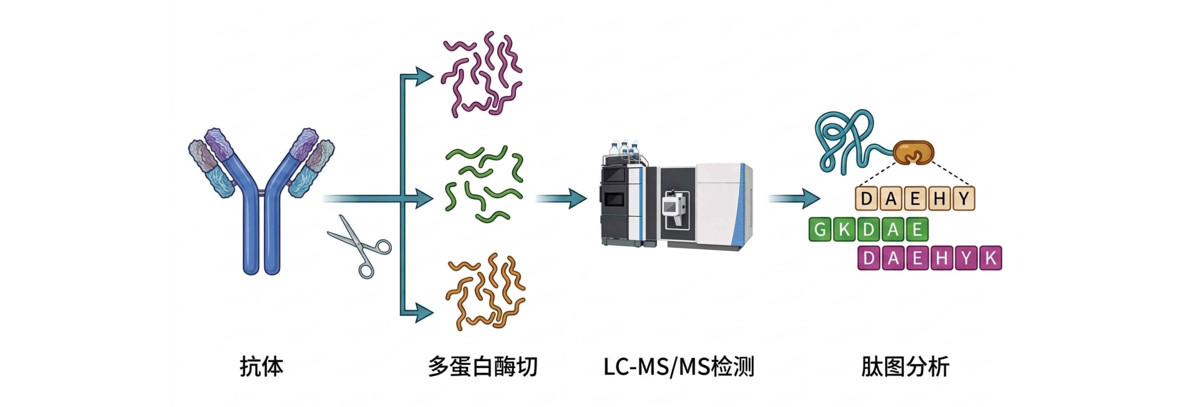
Peptide mapping differs from intact protein molecular weight determination. It involves cleaving proteins into peptides using proteases, followed by LC-MS/MS analysis to align the peptides with the target protein sequence and identify amino acid sequence characteristics. Many critical quality attributes (CQA) analyses of the primary structure of protein drugs are performed using peptide mapping. Peptide mapping analysis allows us to analyze N/C-terminal heterogeneity, confirm protein amino acid sequences and sequence coverage, identify post-translational modification sites and their proportions, and identify sequence variants. Trypsin is the most commonly used protease in peptide mapping analysis; however, single protease digestion rarely achieves 100% sequence coverage. Therefore, mass spectrometry-grade trypsin or multiple protease digestion methods such as Lys-C, Glu-C, and Chymotrypsin are typically used to supplement peptide mapping to achieve 100% sequence coverage.
Analysis Objective
Technical Methods:Peptide Mapping Analysis
Analysis Categories:
Amino acid sequence confirmation
Analysis Objective:
The amino acid sequence of antibody drugs is the material basis for their biological activity and clinical efficacy. After recombinant antibodies are digested by proteases (Lys-C, Asp-N or Glu-C), the amino acid sequence is determined by peptide mass spectrometry using tandem liquid chromatography-mass spectrometry.
Analysis Categories:
N- and C-terminal variants
Analysis Objective:
N-terminal sequencing is an important method for identifying the N-terminal primary structure of antibody drugs. After reduction, the number of N-terminal amino acids in the light and heavy chains of the antibody is determined sequentially by Edman degradation, which can be used as an identification item for batch release testing of the drug substance. The N/C-terminal sequence can also be analyzed in conjunction with amino acid coverage structure to confirm that the terminal sequence is consistent with the target sequence. Due to degradation by carboxypeptidase D in engineered cells, recombinant antibody drugs may also experience incomplete cleavage of the C-terminal lysine residues in the heavy chain. Currently, C-terminal heterogeneity has been confirmed in recombinant antibodies, but there is no evidence that N/C-terminal heterogeneity affects the safety or efficacy of antibodies. However, analysis of N/C heterogeneity helps to strengthen the quality control of recombinant antibody drugs.
Analysis Categories:
Amino acid variation
Analysis Objective:
Changes in the primary structure may also occur in biosimilars of antibody fusion proteins and in the early screening stages of biosimilar development. By combining multiple enzyme digestion peptide mapping analyses, 100% sequence coverage can be achieved, confirming the antibody amino acid sequence.
Analysis Categories:
Post-translational modifications of amino acids
Analysis Objective:
Peptide mapping analysis can precisely analyze the types and proportions of amino acid modifications, such as deamidation, methionine oxidation, glycosylation, N-terminal pyroglutamic acid cyclization, and C-terminal lysine cleavage. Methionine (Met) and tryptophan (Trp) residues are prone to oxidative modification, which may affect antibody stability and function. Deamidation is one of the most common degradation pathways encountered by monoclonal antibody drugs, especially the deamidation of asparagine (Asn) residues in the CDR region. Deamidation is prone to occur when Asn is followed by a small and active glycine (Gly) residue (NG motif). If it occurs in the CDR region, it will lead to a decrease in affinity for antigen binding and loss of antibody potency.
Analysis Categories:
N/O-glycosylation modification (intact glycopeptides)
Analysis Objective:
Glycosylation modification is an important part of the molecular structure of antibody drugs. The N-sugar modification of the antibody occurs at the Asn site in the "Asn Glycosylation modifications play an important role in maintaining the normal structure and biological activity of antibodies, and their impact on function is multifaceted. High-galactose modifications can enhance the CDC effect of antibodies, while low-fucosylation modifications can enhance the ADCC and ADCP effects of antibodies. Non-human glycosylation modifications, such as α1,3 galactose or NGNA, may cause immunogenic reactions and affect the safety and effectiveness of drugs.
Analysis Categories:
Disulfide bonds
Analysis Objective:
The structural features of disulfide bonds have a significant impact on the stability, conformation, and function of antibody drugs. Analysis of the secondary structure of antibody drugs can provide in-depth understanding of their molecular assembly and structural integrity, offering crucial information for quality control. Disulfide bond analysis includes the formation of free thiol groups, thioethers, cysteine oxidation, and trisulfide bonds.
Analysis Process
Key Deliverables
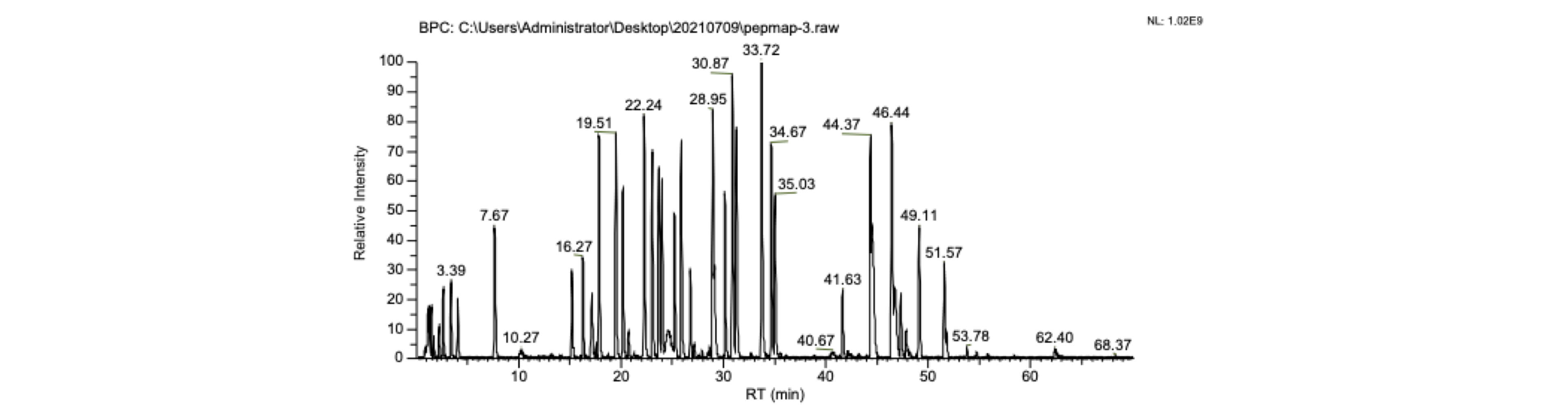
LC-MS/MS peptide map and total ion current peak map
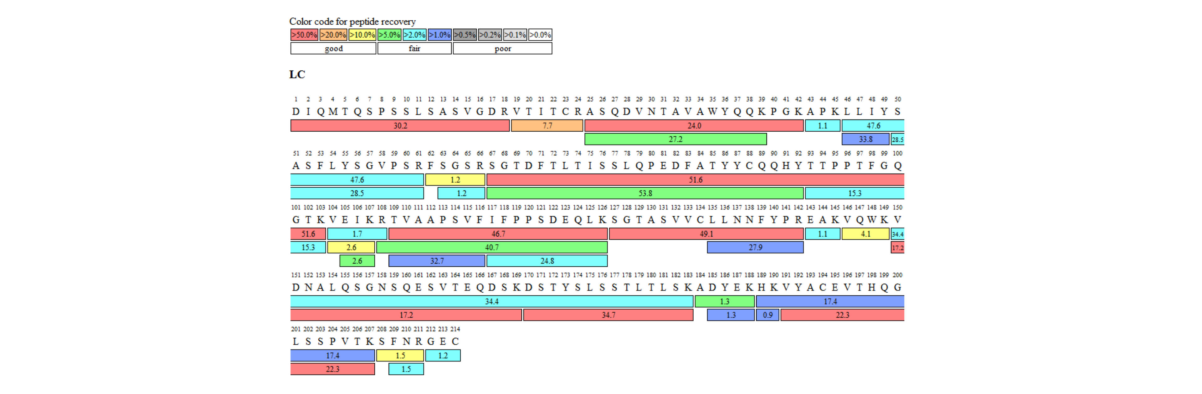 Peptide mapping analysis confirmed amino acid sequence coverage results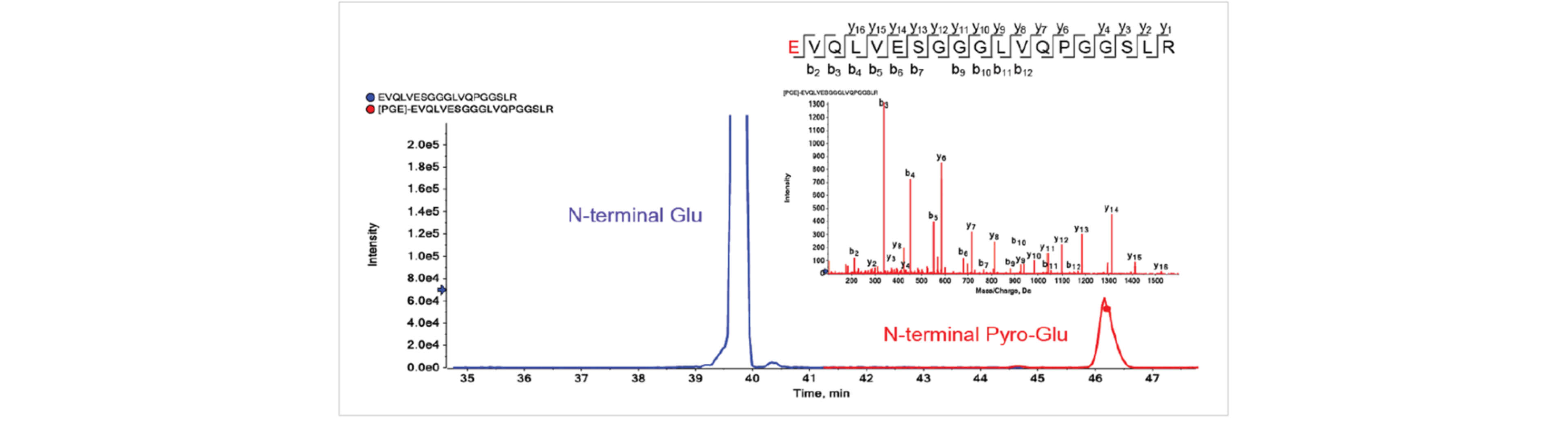
N-terminal pyroglutamic acid cyclization modification of antibody heavy chain
| Protein | Residue # | Modification sites | Category | Sequence | %Abundance |
|---|---|---|---|---|---|
| HC | 55 | N55+Deamidation | Deamidation | IYPTNGYTR | 44.38 |
| HC | 318 | N318+Deamidation | Deamidation | VVSVLTVLHQDWLNGK | 26.68 |
| LC | 30 | N30+Deamidation | Deamidation | ASQDVNTAVAWYQQKPGK | 12.56 |
| HC | 387 | N387+Deamidation | Deamidation | GFYPSDIAVEWESNGQPENNYK | 7.42 |
| HC | 392 | N392+Deamidation | Deamidation | GFYPSDIAVEWESNGQPENNYK | 5.81 |
| HC | 217 | K217+Gln->Pyro-Glu | Gln->Pyro-Glu | KVEPK | 5.86 |
| HC | 374 | G374+Gln->Pyro-Glu | Gln->Pyro-Glu | GFYPSDIAVEWESNGQPENNYK | 1.46 |
| LC | 103 | K103+Glycation | Glycation | SGTDFTLTISSLQPEDFATYYCQQHY TTPPTFGQGTKVEIK | 1.1 |
| LC | 149 | K149+Glycation | Glycation | VQWKVDNALQSGNSQESVTEQDSK | 0.85 |
| HC | 329 | K329+Glycation | Glycation | VSNKALPAPIEK | 0.66 |
| HC | 255 | M255+Oxidation | Oxidation | DTLMISR | 3.67 |
| HC | 107 | M107+Oxidation | Oxidation | WGGDGFYAMDYWGQGTLVTVSSASTK | 1.88 |
| HC | 431 | M431+Oxidation | Oxidation | WQQGNVFSCSVMHEALHNHYTQK | 1.35 |
| HC | 83 | M83+Oxidation | Oxidation | NTAYLQMNSLR | 0.72 |
| LC | 4 | M4+Oxidation | Oxidation | DIQMTQSPSSLSASVGDR | 0.63 |
| HC | 39 | Q39+Gln->Pyro-Glu | Gln->Pyro-Glu | QAPGK | 3.26 |
| HC | 1 | E1-18.0106 | Gln->Pyro-Glu | EVQLVESGGGLVQPGGSLR | 3.69 |
| HC | 249 | K249+Glycation | Glycation | THTCPPCPAPELLGGPSVFLFPPKPK | 0.64 |
| HC | 300 | N300+A2G0F | N-Glycan | EEQYNSTYR | 38.42 |
| HC | 300 | N300+A2G1F | N-Glycan | EEQYNSTYR | 36.11 |
| HC | 300 | N300+A1G0F | N-Glycan | EEQYNSTYR | 9.7 |
| HC | 300 | N300+A2G2F | N-Glycan | EEQYNSTYR | 6.97 |
| HC | 300 | N300+A2G0 | N-Glycan | EEQYNSTYR | 3.37 |
| HC | 300 | N300+A1G1F | N-Glycan | EEQYNSTYR | 3.24 |
| HC | 300 | N300+A2G1 | N-Glycan | EEQYNSTYR | 1.85 |
Peptide post-translational modification ratio

Shenzhen Wininnovate Bio Co., Ltd.
Innovative mass spectrometry and AI technologies provide protein and metabolite mass spectrometry multi-omics solutions for life science research, empowering the growth of the biotechnology, pharmaceutical, and healthcare industries.


